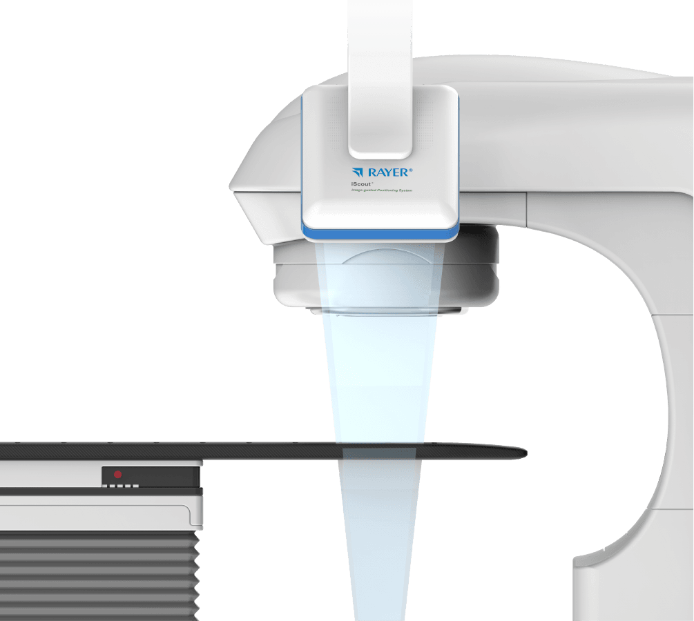
iSCOUT
Image-guided radiotherapy positioning system
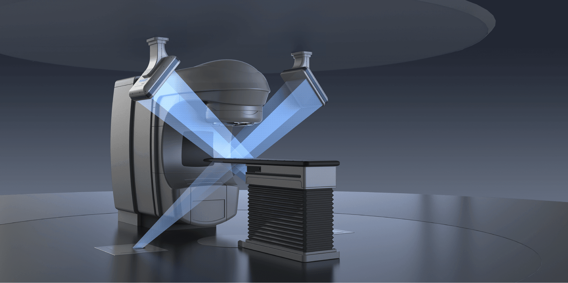
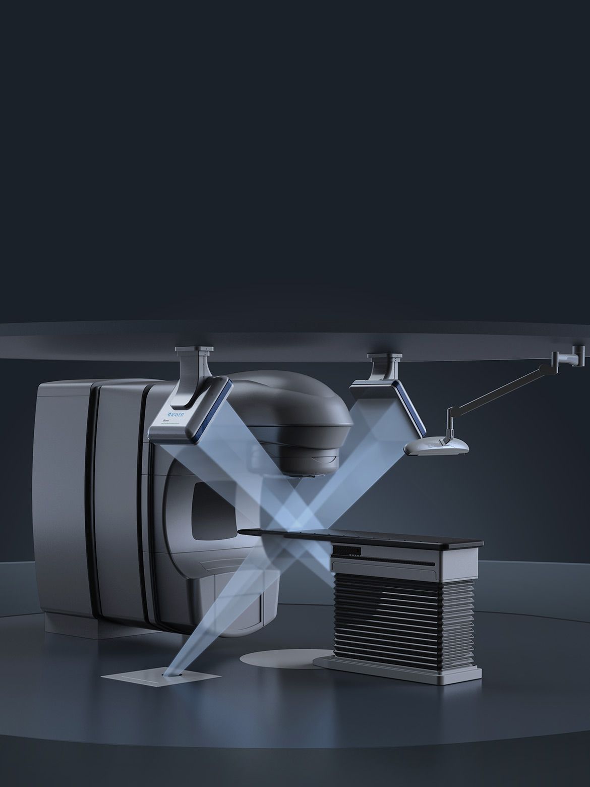
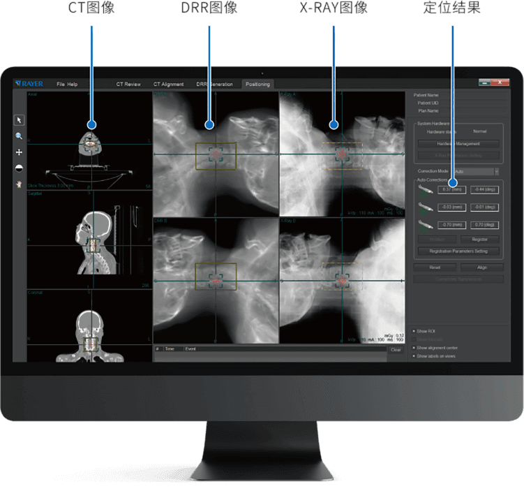
Online Image Guidance & Real-Time Position Verification
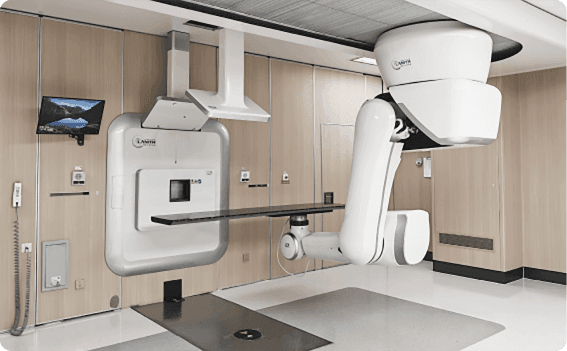
Using kV level X-ray stereoscopic planar imaging technology, the product software controls two sets of X-ray imaging units that are configured symmetrically to positioning center, simultaneously obtaining two orthogonal projection X-ray images. Then Image registration is performed by using the anatomical structure features to realize the patient positioning verification. The product is combined with radiotherapy equipment to detect and correct the patient setup errors and verify the final residual setup errors before radiation treatment.
-

Precise
Positioning accuracy less than 1.0mm
-

Fast
Positioning time: 1-3 minutes
-

Convenient
Remote one-click couch movement to streamline workflow
-

Real-time
Supports intrafractional position verification
-

Low dose
Head: 0.01-0.03 cGy Chest: 0.03-0.08 cGy Pelvis: 0.04-0.11 cGy

Large-Field Imaging Coverage
Effective imaging area of 434 mm × 434 mm, expanding anatomical structure imaging range Image resolution up to 2816 × 2816 pixels, enhancing image clarity Dynamic imaging frame rate up to 25 fps
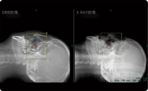
Diagnostic-Grade Orthogonal Image Quality
Flat panel withstands MV-level high-energy therapeutic radiation exposure Shortened X-ray penetration path significantly improves image quality More intuitive and accurate visualization of anatomical structures

Remote Rapid Setup Verification
One-click remote couch movement for rapid setup error correction Re-verification of residual setup errors
Technical Advantages
-

Suitable for whole-body target visualization and positioning
-

Capable of both inter-fraction and intra-fraction setup verification
-

Detects setup errors in 3 translations and 3 rotations
-

Positioning accuracy better than 1.0 mm
-

Short positioning time with low imaging dose
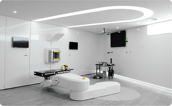
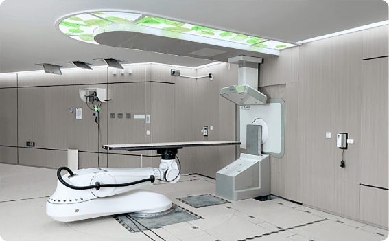
Combined with Multiple Models of Radiotherapy Equipment

Independent installation
No mechanical/electrical connections to radiotherapy equipment

Short installation time
No interference with treatment room operations

Device portability
Flexibly adapts to hospital clinical requirements
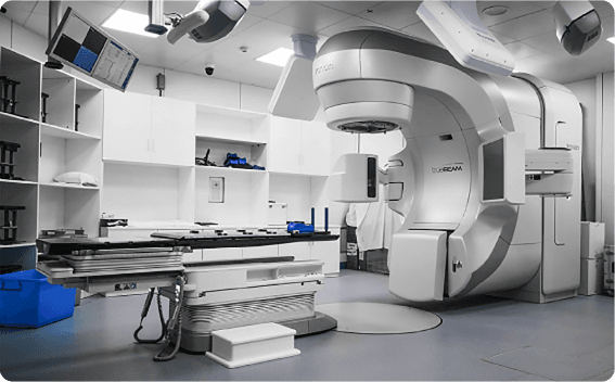
Linear Accelerator Radiotherapy System
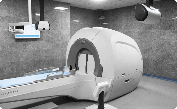
Gamma Ray SRS System
Gamma Ray SBRT System
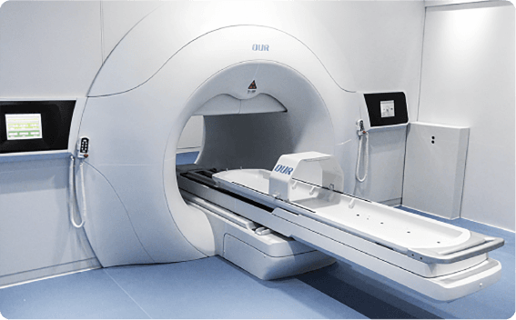
Proton Accelerator System
Heavy ion Accelerator Configuration
Boron Neutron Accelerator
iSCOUT、CBCT contrast
| Product name | iSCOUT | CBCT |
| Working principle | kV-level stereoscopic planar imaging | 3D imaging |
| Structure features | Double detectors, double tubes, double generators | Single detector, single tube, single generator |
| Image quality | kV-level | kV-level or MV-level |
| Imaging method | Instant image capture | Continuous rotation imaging |
| Positioning time | Within 1 minute | 2~3min |
| Imaging dose | 0.2-1.0 mGy | 30-80 mGy |
| Usage frequency | Interfraction and intrafraction | Interfraction |
| Installation method | Independent installation, no mechanical and electrical connections | Integrated installation (weight matching, mechanical and electrical connections) |